CASE 8: Revision of a metal-on-metal hip for adverse reaction to metal debris (ARMD) using a bearing exchange

The Story
“Gail was having awful pain in her right hip which had been worsening for 4 years - to the point where she was using a walking stick to mobilise. Bilateral metal-on-metal Pinnacle S-ROM total hip replacements had been in situ for 8-years on the right and 6-years on the left.
For Gail, the greatest challenge was working out the source of her pain. We had to consider loosening, an infection and an adverse reaction to metal debris. This required a detailed work-up.”
The Investigation:
On clinical examination:
Very painful right hip movements
Reduced range of movement on the right
Able to straight leg raise on the right but this caused considerable discomfort
Her oxford hip scores were 17/48 and 29/48 for the right and left hips respectively.
Blood inflammatory markers demonstrated a normal ESR (26mm/Hr) and a borderline CRP (10mg/L). Blood metal ion levels demonstrated a normal level (for a patient with bilateral metal on metal hip implants) of cobalt (2.8ppb) and chromium (3.5ppb). Greater than or equal to 7ppb of either cobalt or chromium is defined as concerning by the MHRA (for a unilateral MOM hip). Due to her borderline raised inflammatory markers, an aspiration of the right hip was performed however no growth was seen after extended cultures, so infection was unlikely.
The Evidence

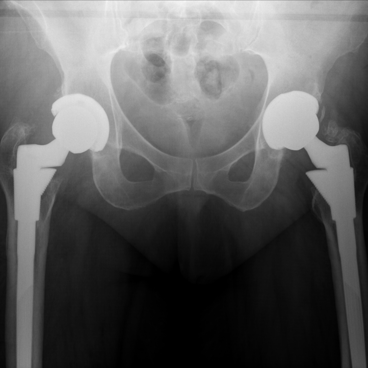
Anteroposterior plain radiograph of the pelvis demonstrates bilateral well fixed and well positioned S-ROM Pinnacle MOM hip replacements. Both cups and stems appear stable and well positioned.

Metal artefact reduction sequence (MARS) MRI demonstrating a large pseudotumour posterior to her right hip. This is an area of inflammation secondary to metal debris which can mimic an infective process in the absence of infection (including raised inflammatory markers, despite no infection in some cases).
The Diagnosis
Gail had an adverse reaction to metal debris (ARMD) as a result of the metal on metal (MOM) bearing in used in her right hip replacement.
The Plan
In the presence of the well fixed Pinnacle cup and SROM stem on the right side, we planned to perform an isolated liner exchange, changing the bearing from metal-on-metal to ceramic-on-polyethylene. As part of the same operation, we aimed to excise Gail’s pseudotumour, as fully as possible (but inevitably some will be left behind).
The Operation
We used a posterior approach to access the right hip and the pseudotumour. We excised as much of the joint lining and the pseudotumour as possible. As with all of our procedures where a possible infection may be the cause, we sent 5 samples of the hip capsule for extended microbiological culture.
We then extracted the femoral head. With an SROM stem this is normally a straightforward task. With other stems, there can be cold welding of the stem to the femoral head (such as the Biomet Magnum head and Taperloc stem) in which case, stem extraction is required. The metal liner was then extracted from the Pinnacle cup using a Depuy “alternate bearing extractor” but it is also possible to extract the liner using a sucker and two simultaneous cup impactors that repeatedly impact the cup rim and vibrate the liner loose from the shell.
We completed the procedure by inserting a 52mm polyethylene liner and a 36mm ceramic head with a titanium sleeve. The joint was relocated and then washed out before the musculature, fascia and skin were sutured.
The Outcome
Anteroposterior plain radiograph demonstrating the new liner of the right THR. The head bearing surface is visible on the right side because the right acetabular liner is now made of polyethylene.

EOS imaging taken 18-months after Gail’s operation. When compared to the post-operative image, there is no migration of either implant and her legs remain the same length.
The Verdict
“Components such as the acetabular shell and femoral stem, can be retained if they are well fixed to bone, well orientated and not infected.”
-
-
-
Please see our other cases of revision surgery for metal debris disease (due to cobalt-chromium debris arising from metal on metal bearing surfaces, metal on titanium taper junction surfaces, cemented stem surfaces)