CASE 13: Revision of a loose acetabulum with pelvic discontinuity due to polyethlene osteolysis, using a custom 3D printed cup

The Story
“The patient is a 49-year old female with psoriatic arthritis. She has multiple joint replacements including bilateral knee, bilateral elbow and a left hip replacement.
She presented to clinic with pelvic discontinuity. Hip movement was restricted and the leg was short clinically by 3 to 4cm. She had an antalgic gait. In addition, she had pain in the groin running down her left leg.
The initial acetabular component was removed to allow for a planning CT scan to be taken. This involved a vascular surgeon to free the internal iliac arteries from the acetabular metal work. The images below represent the second stage of her hip reconstruction.”
The Evidence
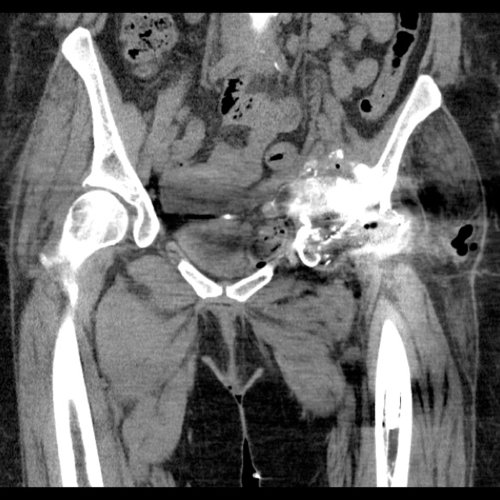
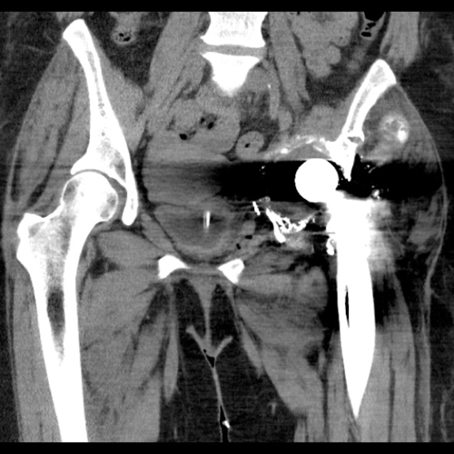
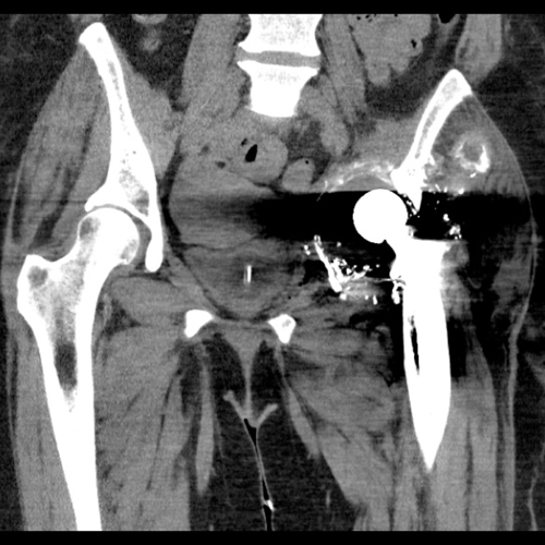
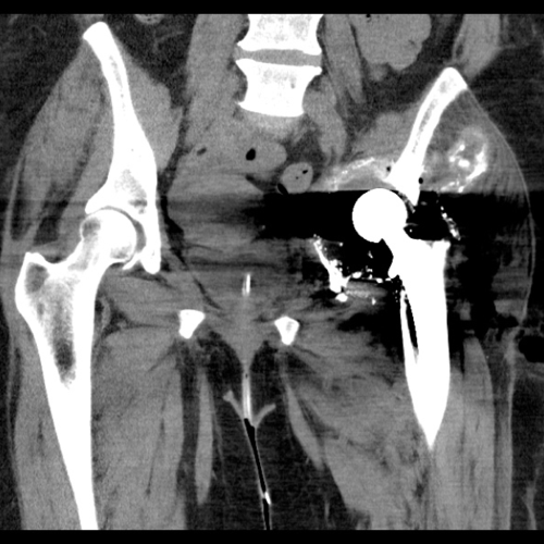
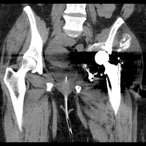
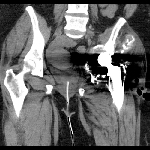
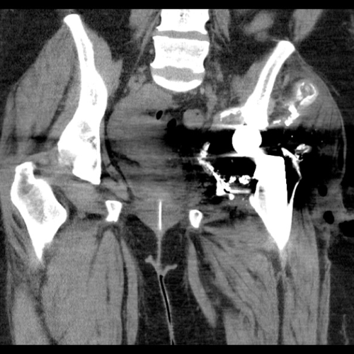
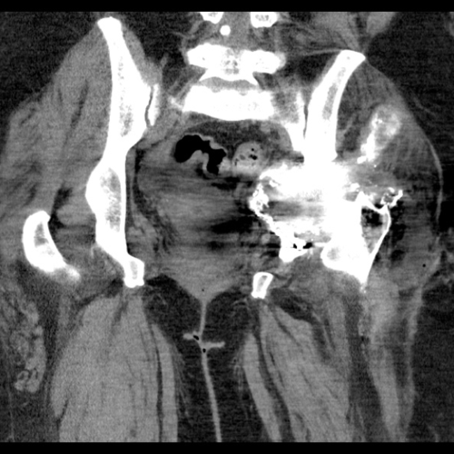

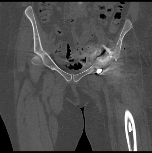
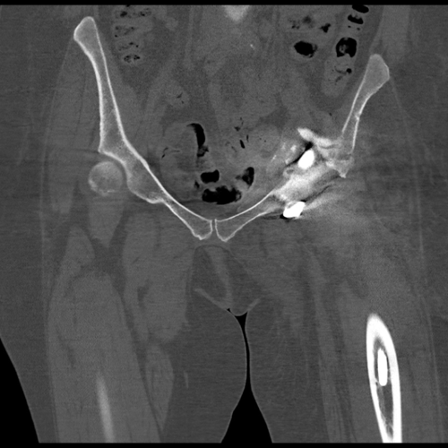
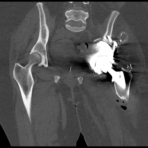
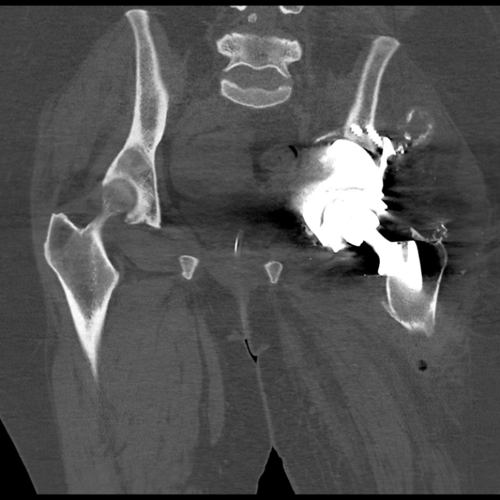
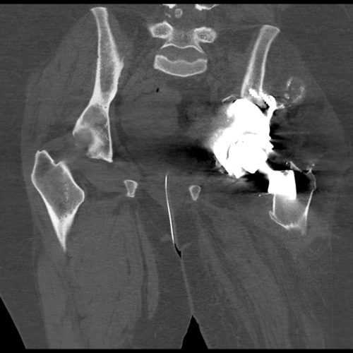
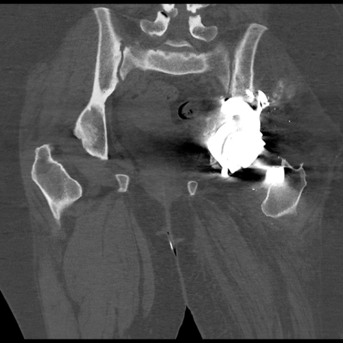
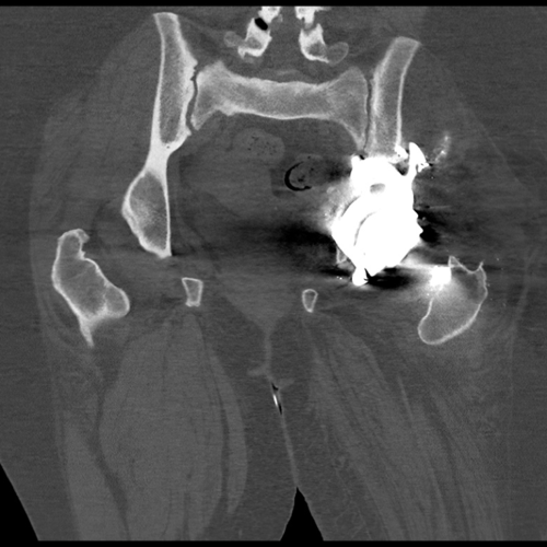
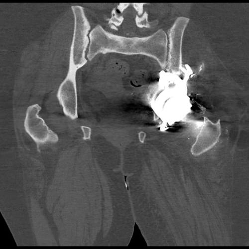
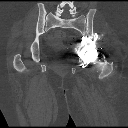
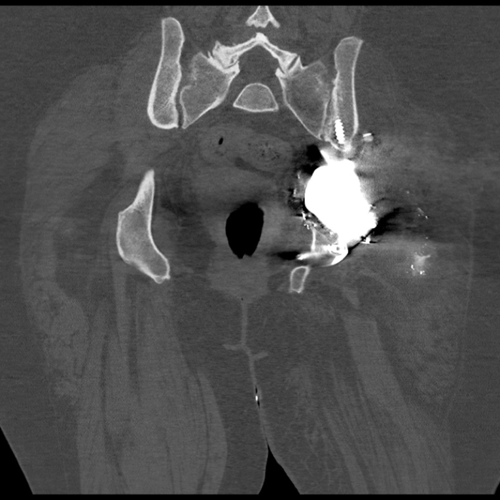
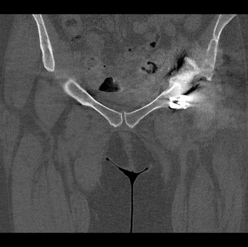
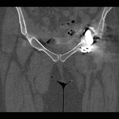
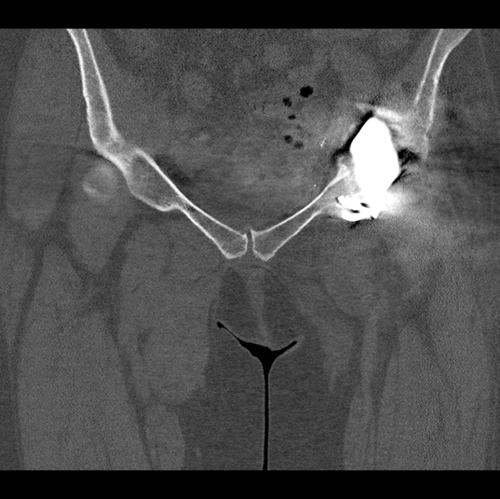
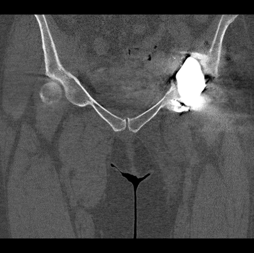
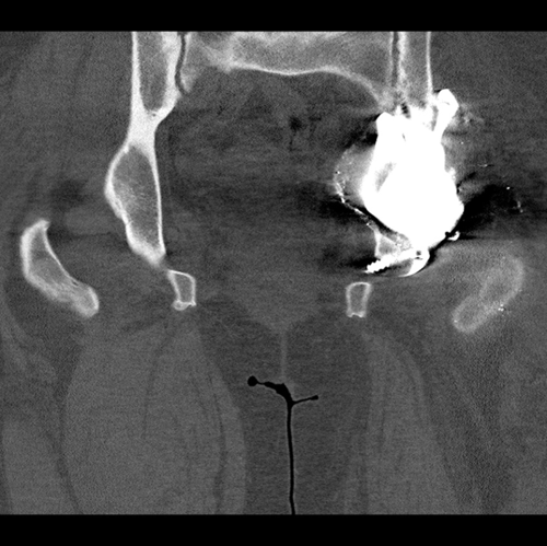
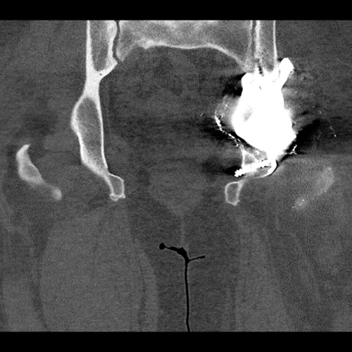
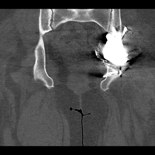
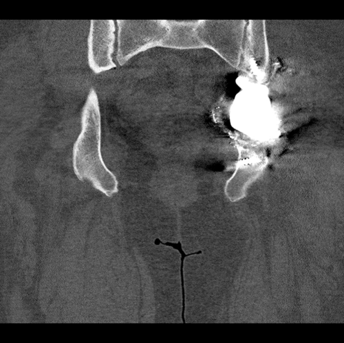
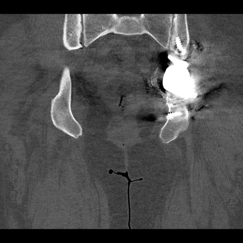
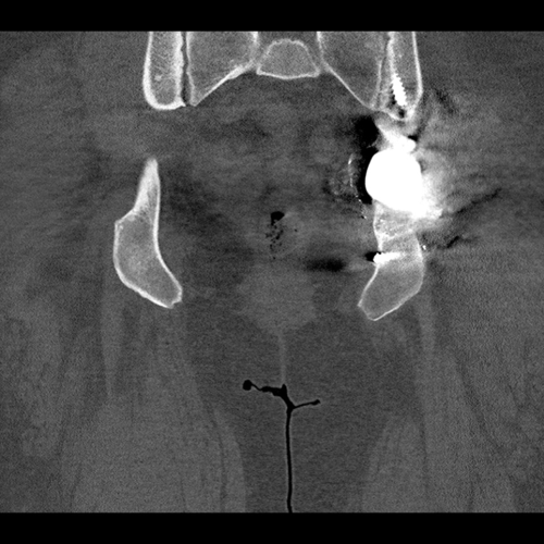
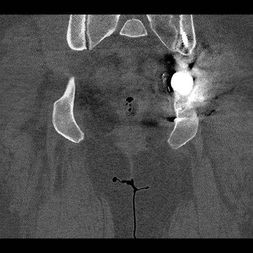
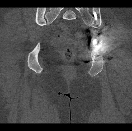
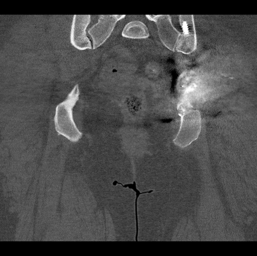
CT scan showing the patient’s pelvis without the acetabular metal work. This was used to make the hemipelvic 3D model seen below.
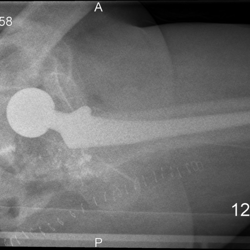
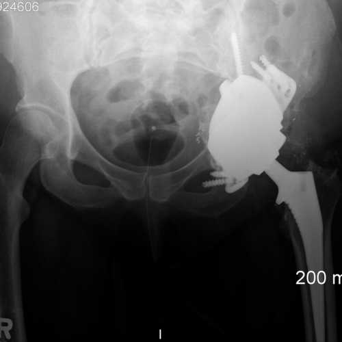
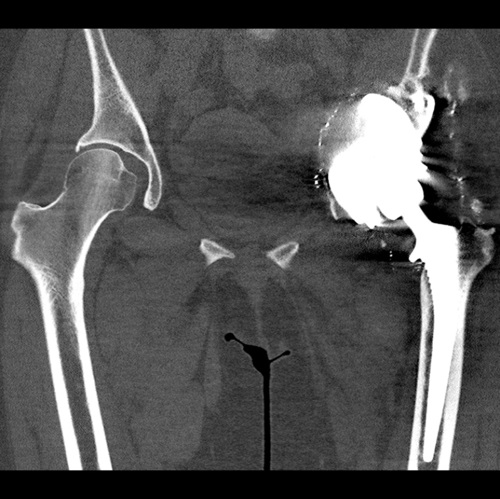
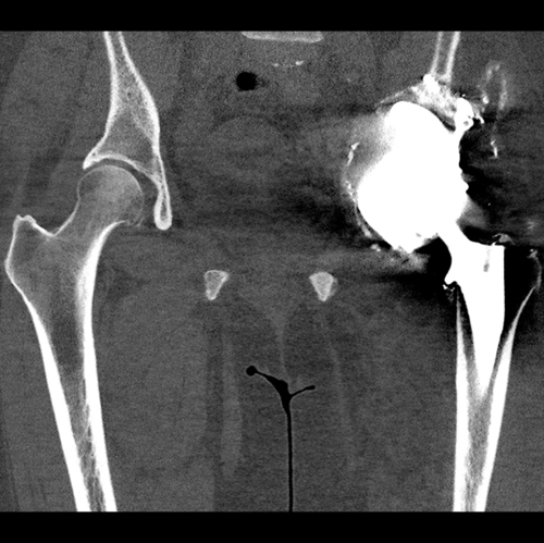
Pre-operative radiographs showing the patient’s left hip after the first stage procedure. The surgical clips are present. Note there are surgical clips in the inguinal region for the ilioinguinal approach to free the intrapelvic vessels and further clips posteriorly to access and remove the acetabular cup.


The 3D hemipelvis model highlights the extent of the bone loss. There is complete pelvic discontinuity.
The Diagnosis
Pelvic discontinuity with a history of psoriatic arthritis. This defect is classified as a Paprosky 3B with pelvic discontinunity.
The Plan
Open reduction internal fixation using a distal femoral locking plate prior to the acetabular reconstruction.
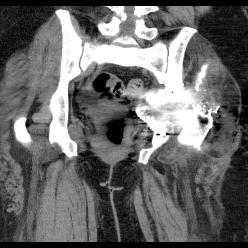
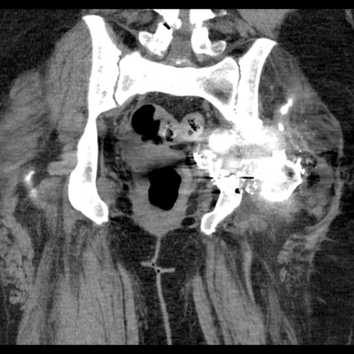
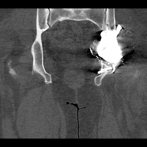
The patient underwent a CT scan after the right femoral fracture to plan her custom acetabular component.
Implant Design
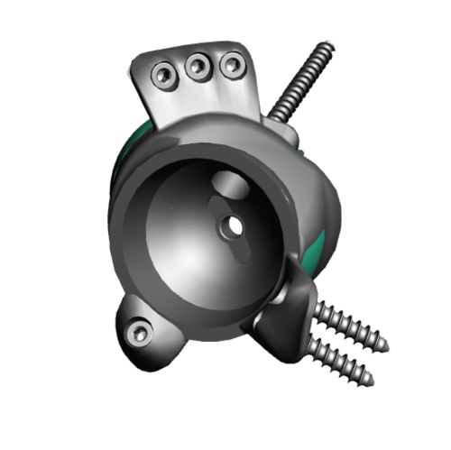
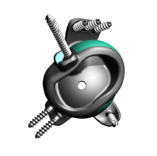
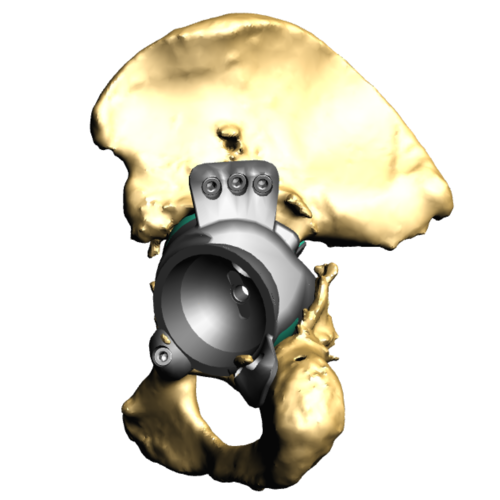
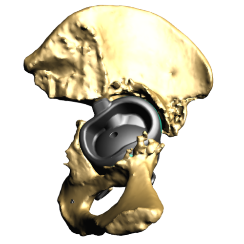
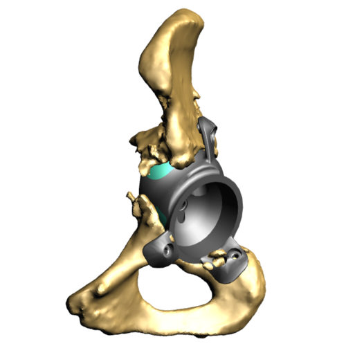
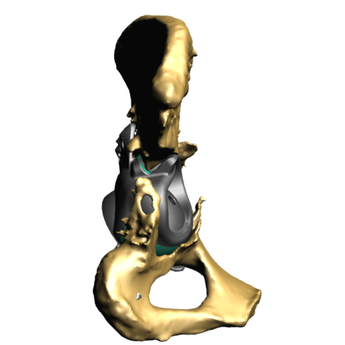
This series of images shows the implant design and images of it within the pelvic 3D model. This implant has been designed to bridge the pelvic discontinuity
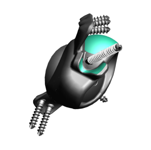
Trabecular Titanium

This image shows the trabecular titanium metallic structure. This is manufactured using 3D printing methods to produce a continuous porous structure to encourage bony osseointegration
The Operation
Key surgical steps:
Posterior approach to allow extensive exposure of acetabulum and femur.
Release of psoas and gluteus maximus from the femur, and abductors from the ilium.
Bone preparation with reamers according to 3D computer plan and checking using sterilised plastic 3D-printed models of the acetabular pre and post bony preparation.
Wash.
The Outcome
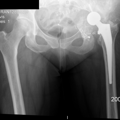
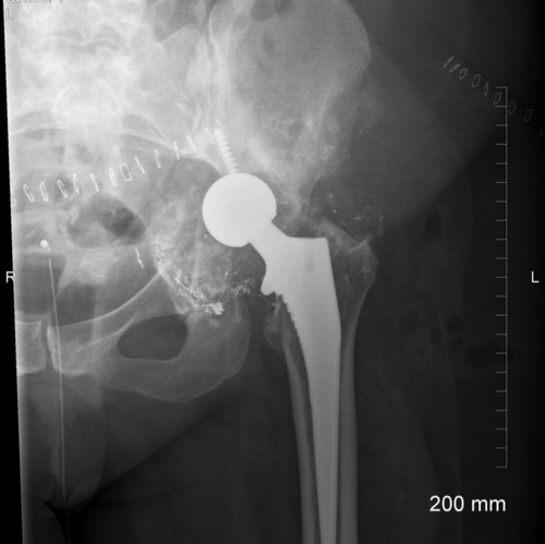
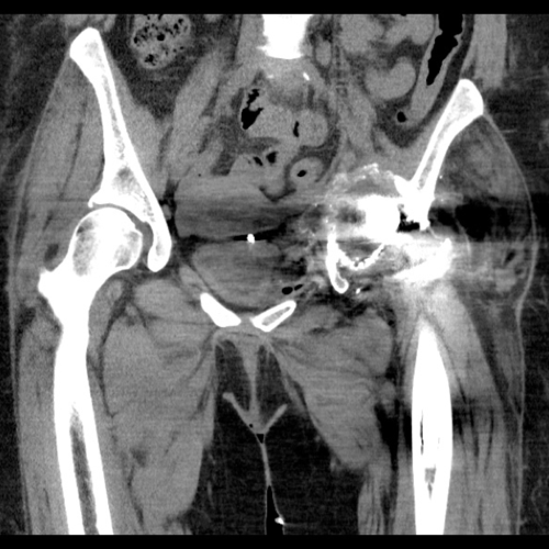
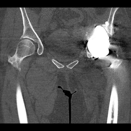
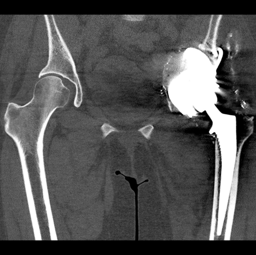
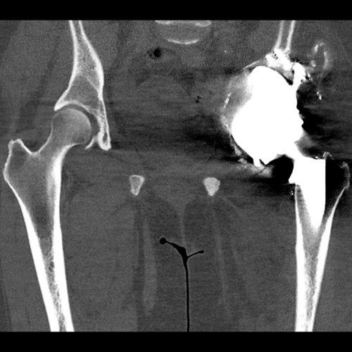
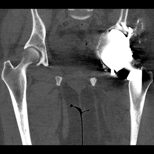
Post-operative images after the second stage of the patient’s procedure. The implant is holding together the superior and inferior aspects of the patients left hemipelvis.
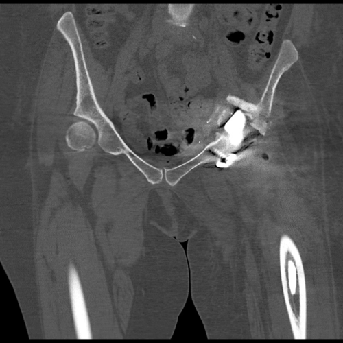
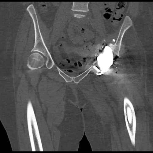
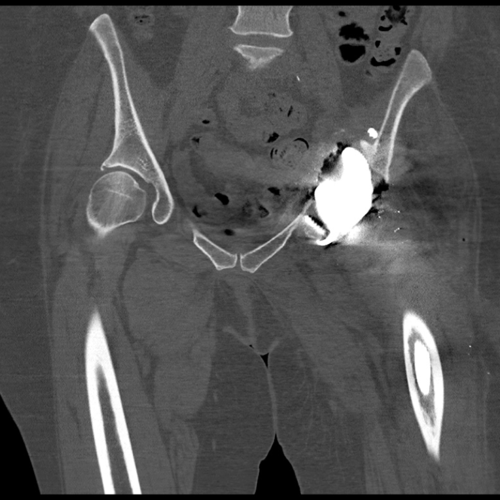
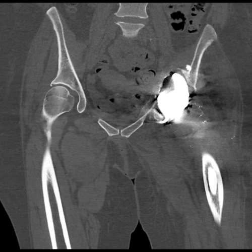
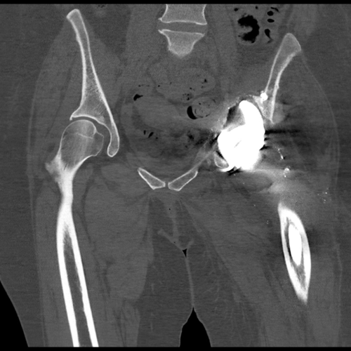

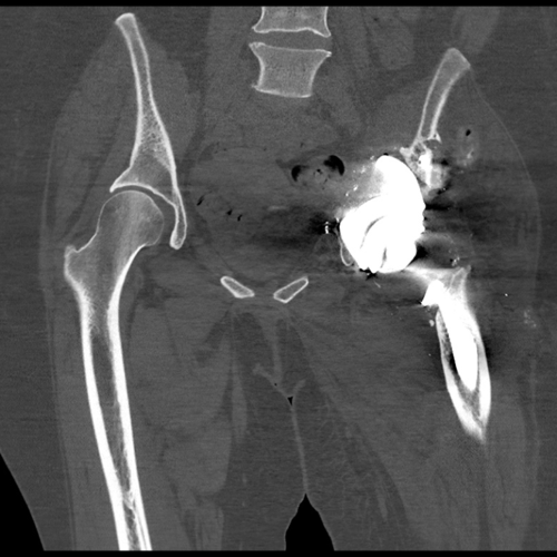
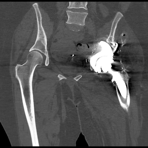


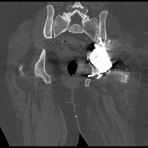


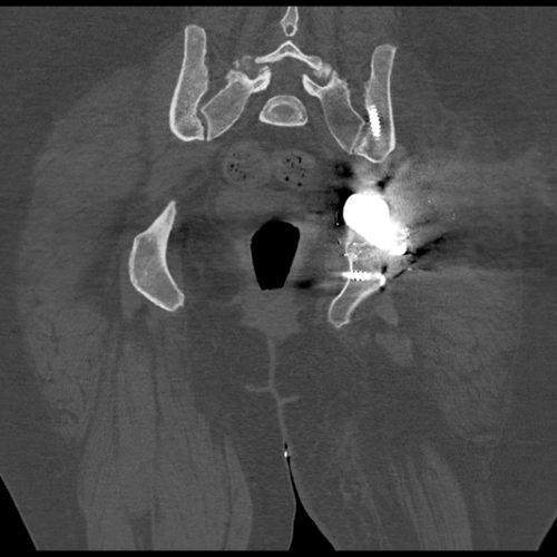
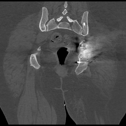
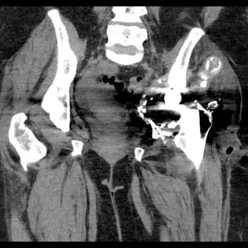
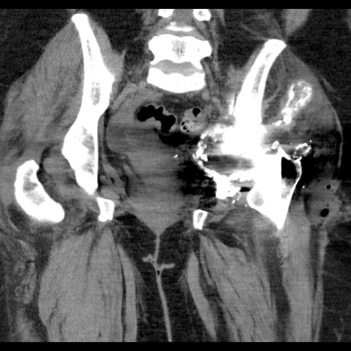
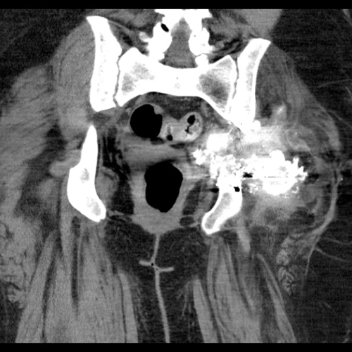
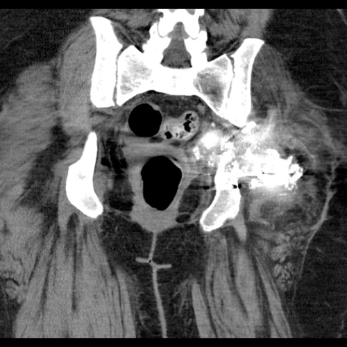
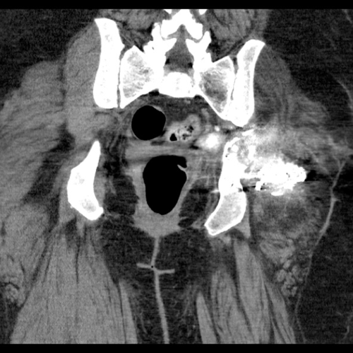
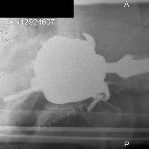
CT scan demonstrates optimal positioning of the acetabular component
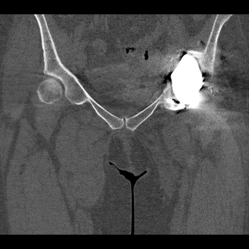
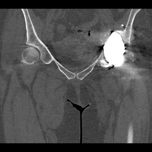
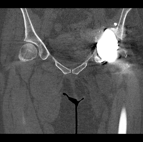

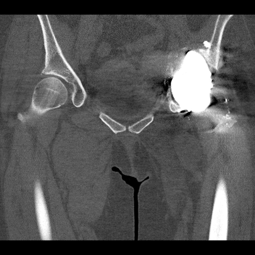
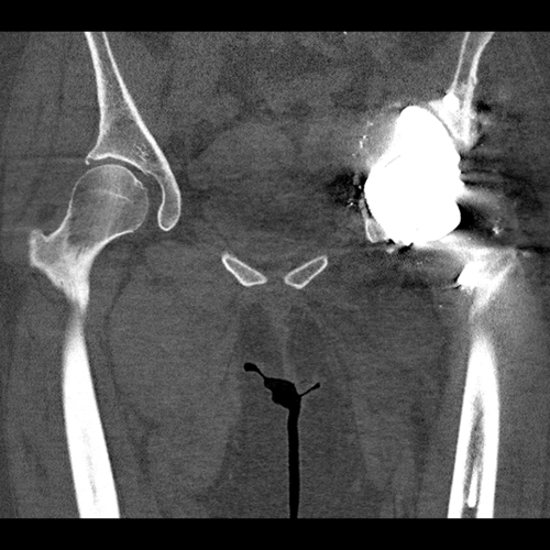
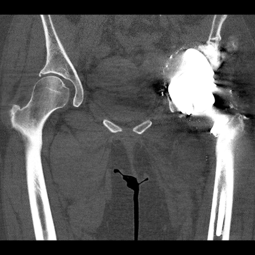
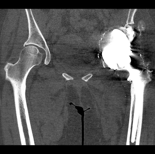
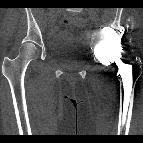
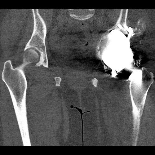
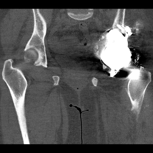
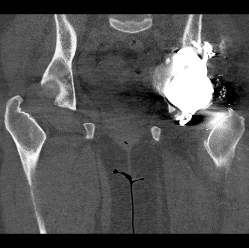
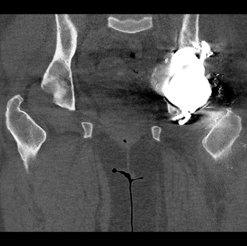
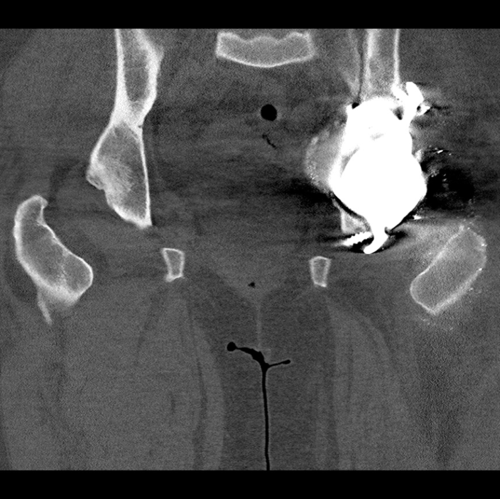
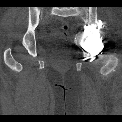
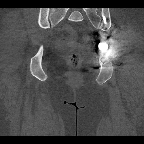
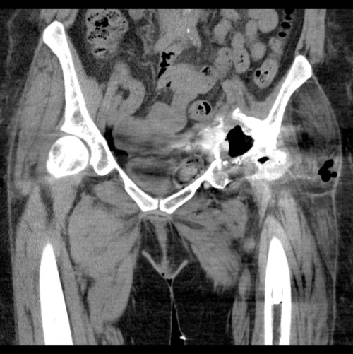
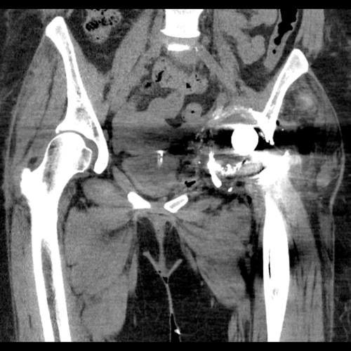
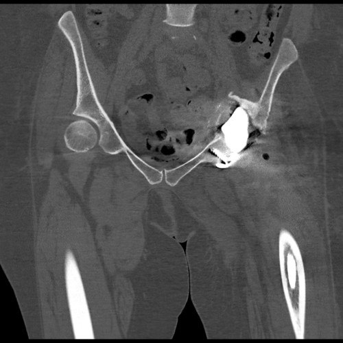
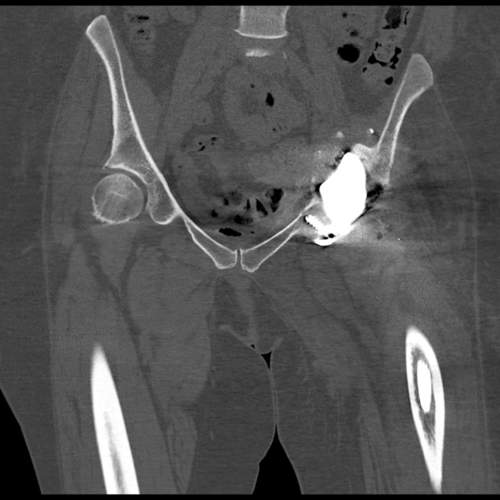
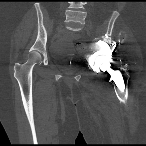
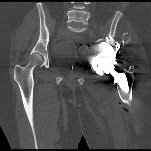
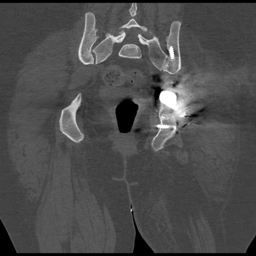
CT scan at 7-months shows no migration of the implant.
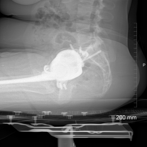
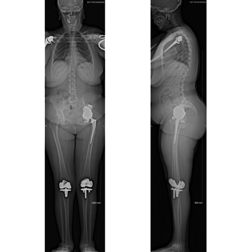
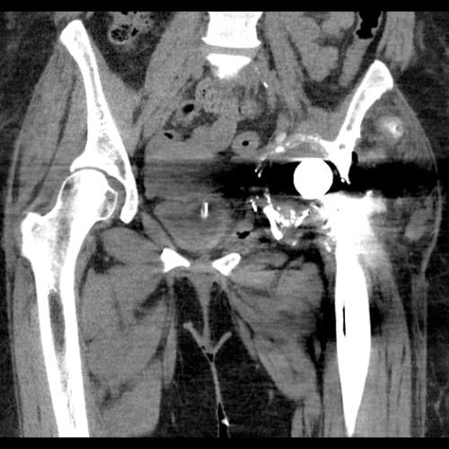
Post-operative EOS images demonstrate the implant in a functional position. There is no functional leg length discrepancy or pelvic tilt.
The Verdict
“At the 1-year follow-up consultation the patient reported that the left sided hip pain was no longer present at rest. She was able to complete activities around the house and was able to sleep without the need of analgesics.
Clinically, the leg length discrepancy has been corrected, and the patient has regained a good range of movement in her left hip.”