CASE 10: Revision of a metal on metal hip with massive acetabular osteolysis and previous femoral osteotomy using a custom 3D-printed cup in a mid-life woman

The Story
“In 2001, at the age of 32 years, Zara underwent metal-on-metal (MoM) hip resurfacing. 17 years later, she presented with mild hip pain but extensive loss of bone in the pelvis due to an inflammatory reaction to the metal wear particles. Her blood metal ion levels were 100 times greater than patients with well functioning MoM hips.
Without surgery, the pelvis was at imminent risk of fracture, which would then need even more complex emergency surgery. However, surgery also risked fracture of the pelvis during removal of the implant.
And since the remaining pelvic bone was poisoned by the metal debris, I didn’t know whether the new implant would in fact fix to the pelvis. All these risks had to be weighed up to ensure the best possible outcome for Zara.”
The Investigation
The 49-year-old presented with mild left hip pain and raised cobalt and chromium levels (Co = 146ppb and Cr = 120ppb). 17 years previously she had undergone a left Birmingham MoM hip resurfacing due to unilateral hip dysplasia (DDH). The commonly used, alternative type of hip replacement – metal-on-polyethylene – was not used as this would have worn through the plastic liner and caused bone loss from an inflammatory reaction to the plastic wear particles.
Zara had an excessively anteverted femur at the time of the resurfacing, leading to dislocation in the follow-up phase. For this she received a subtrochanteric derotational osteotomy using a lateral plate.
Her first set of blood tests at our clinic suggested her metal ion levels were rising with a cobalt of 188ppb and a chromium of 126ppb. She had no known systematic problems (heart, endocrine and brain etc.).
Examination revealed the left leg was slightly shorter than the right. There was a good range of movement throughout on both sides, but there was some clunking with rotation of the left leg.
At the time of referral, Zara was still able to ski, do her gardening and walk a few miles.
The Evidence
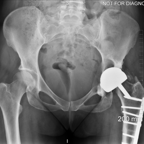
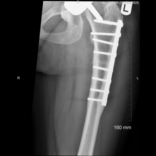
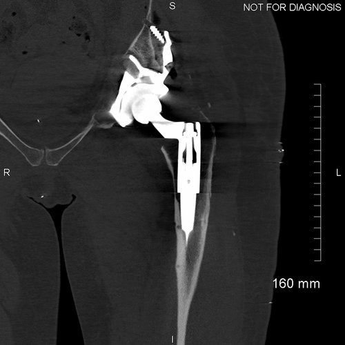
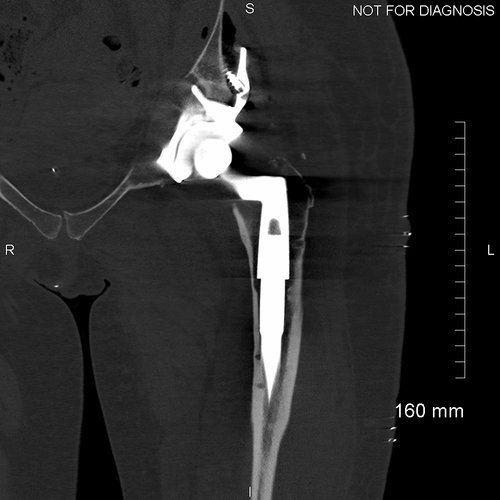
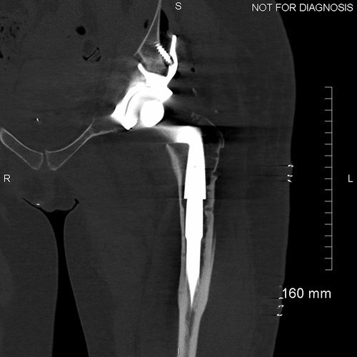
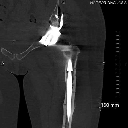
Anteroposterior and lateral plain radiographs demonstrate radiographic features of osteolysis around the acetabular and femoral components. Zara was asymptomatic.

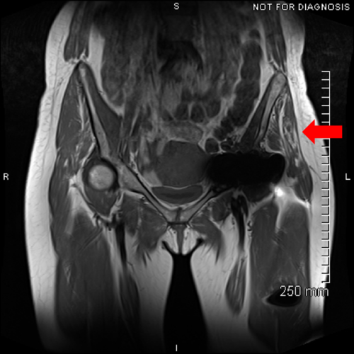
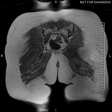
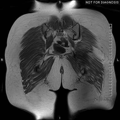
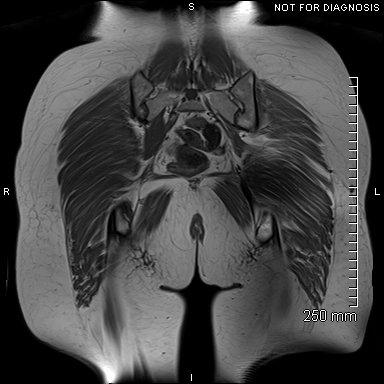
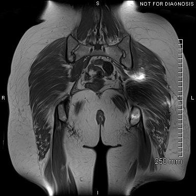
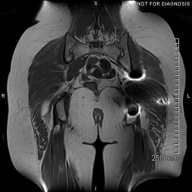
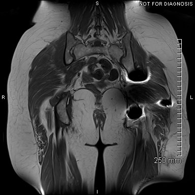
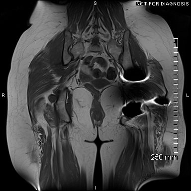
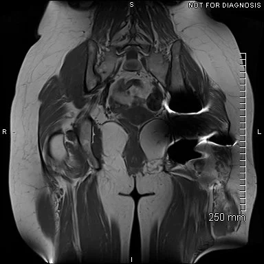
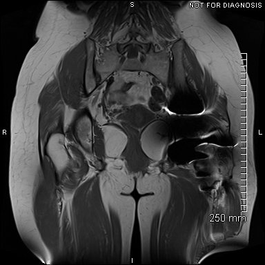
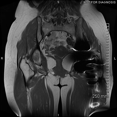
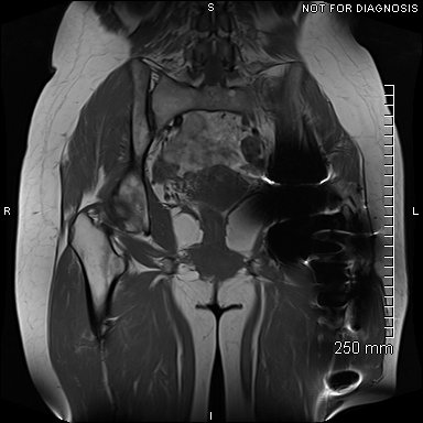
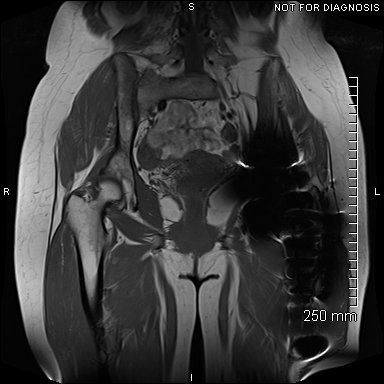
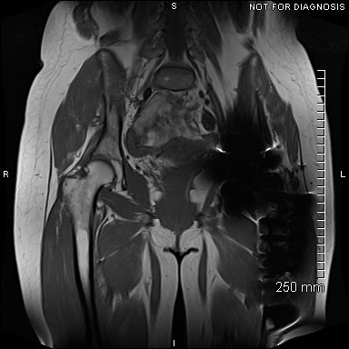
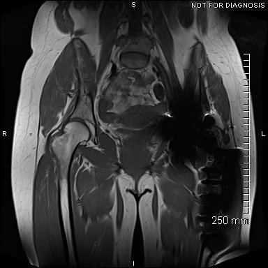
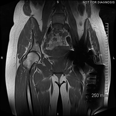
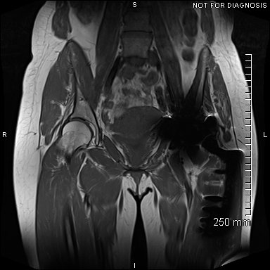
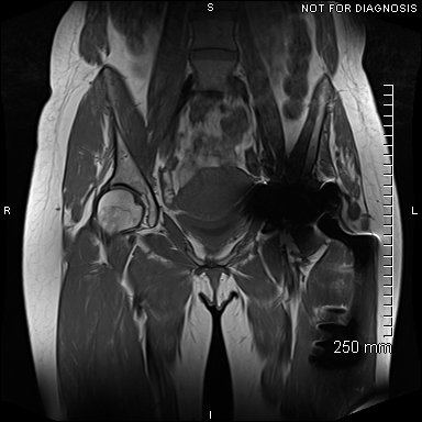
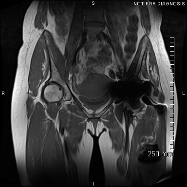
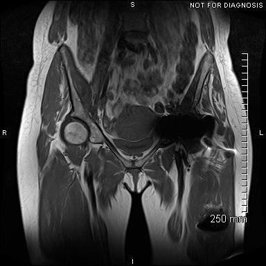
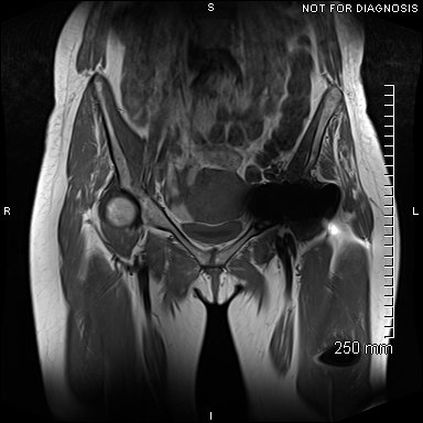
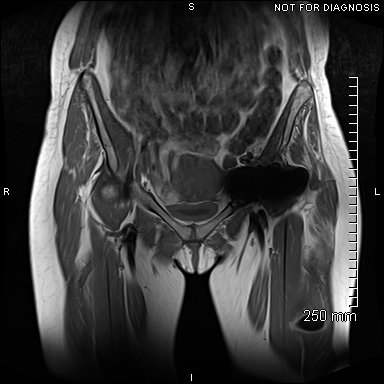
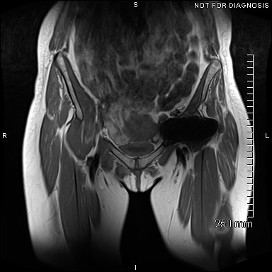
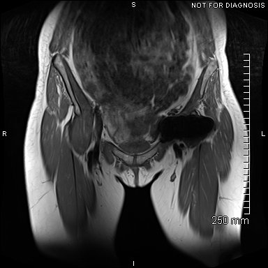
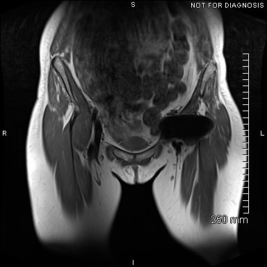
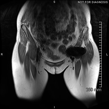
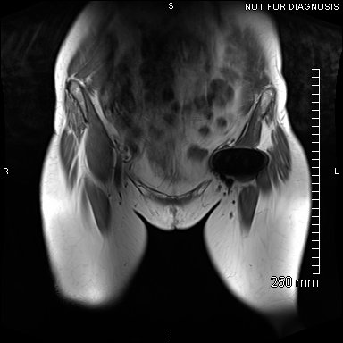
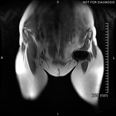
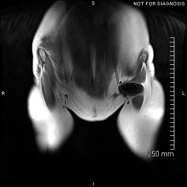
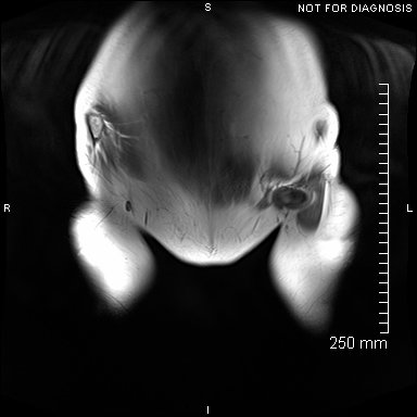
Coronal MRI showing left sided gluteal muscle wasting but no signs of loosening. There were no other abnormalities.

EOS scanning (low radiation dose, 3D scanning in functional positions of standing and sitting) was used to assess leg lengths whilst standing and pelvic tilt whilst sitting.
The Diagnosis
A multi-disciplinary team (MDT) meeting concluded that revision was required due to very raised blood metal ion levels (MHRA suggest levels above 7ppb are of concern) and imminent risk of peri-prosthetic acetabular fracture. They suggested a further CT scan to assess the remaining bone stock.
The Plan
Our plan was to design and fit a custom 3D-printed acetabular implant. We would have to take great care not to cause any fracture during the removal of the MoM hip surfacing as the new implant would only work if the pelvis was intact.
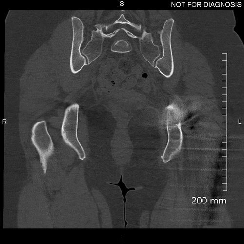
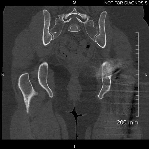
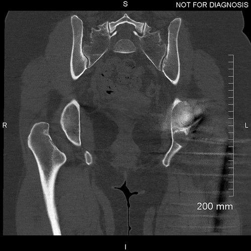

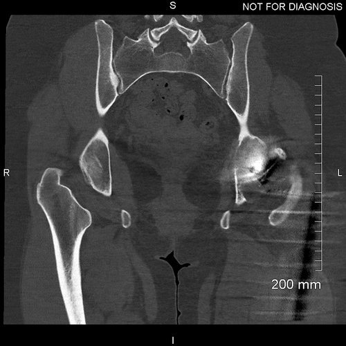
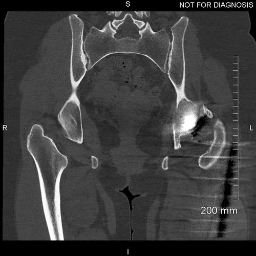
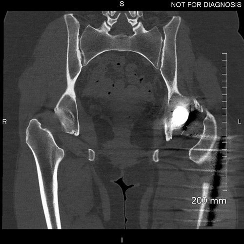
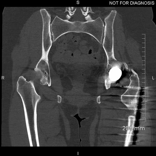
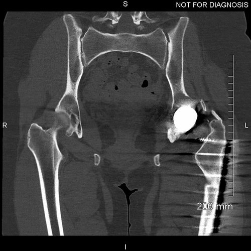

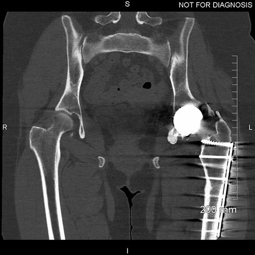
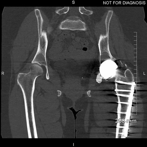
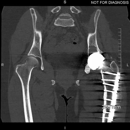
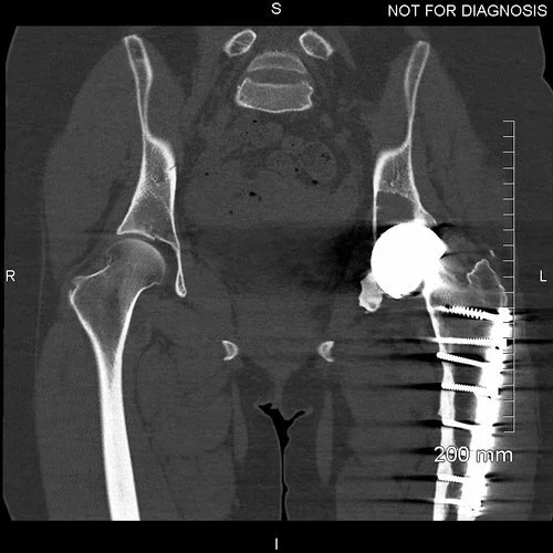
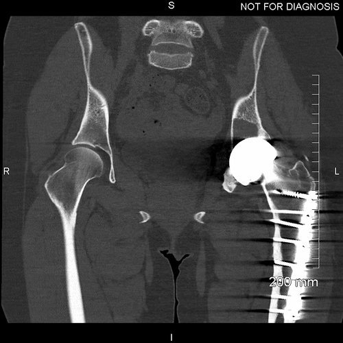

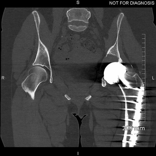

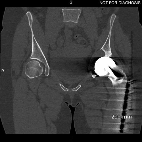
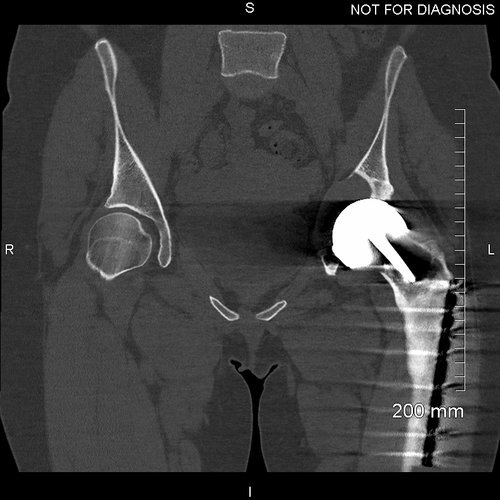

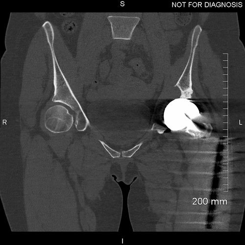
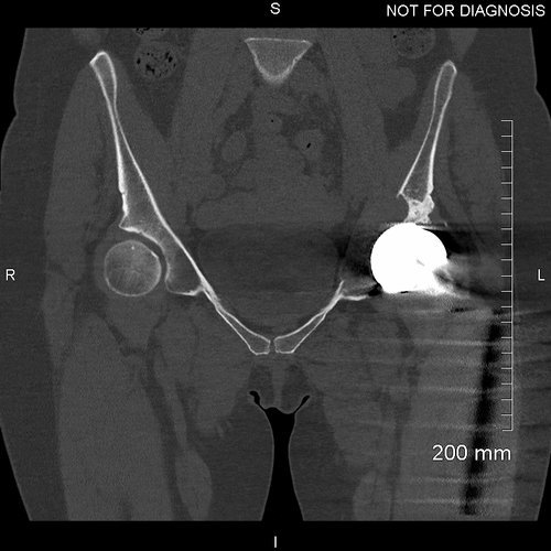

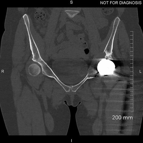
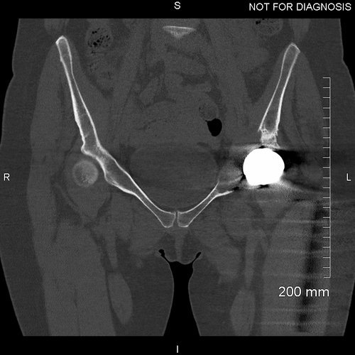
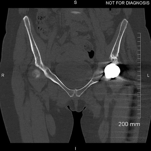
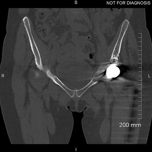
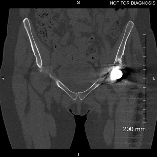
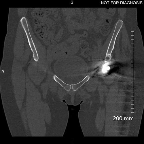
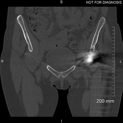
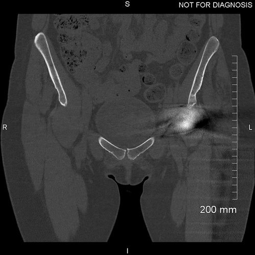
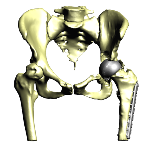
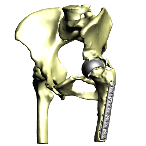
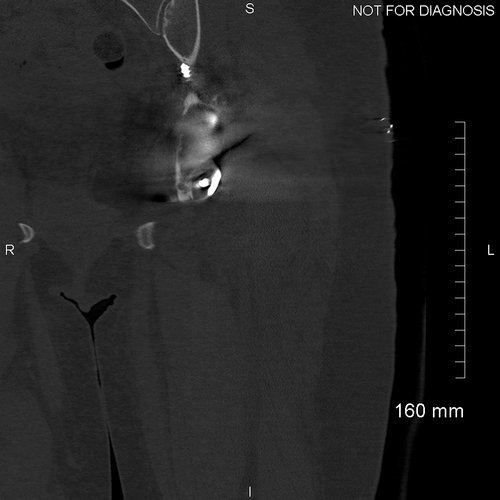
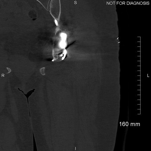
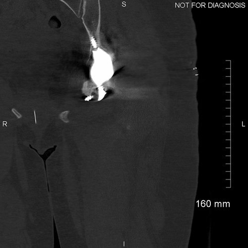
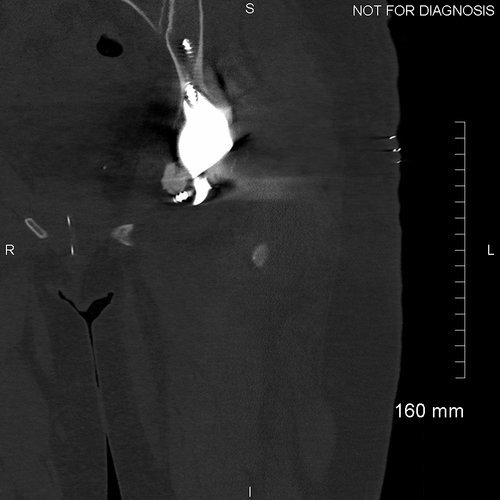
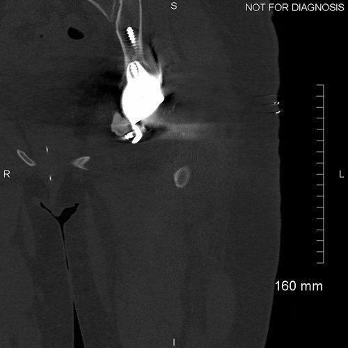
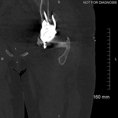
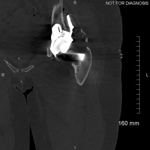
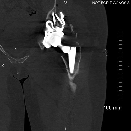
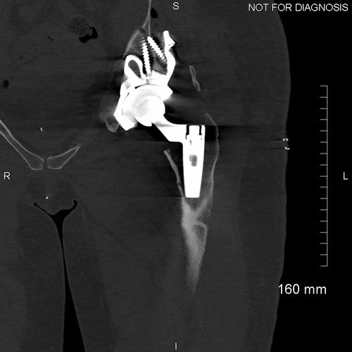
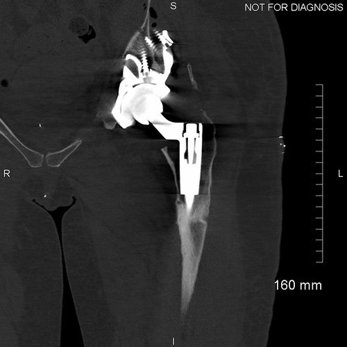
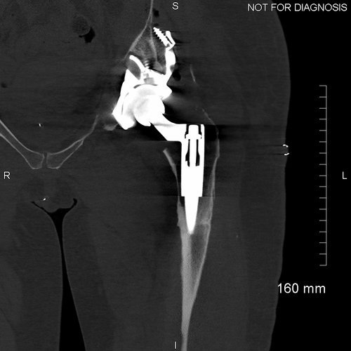
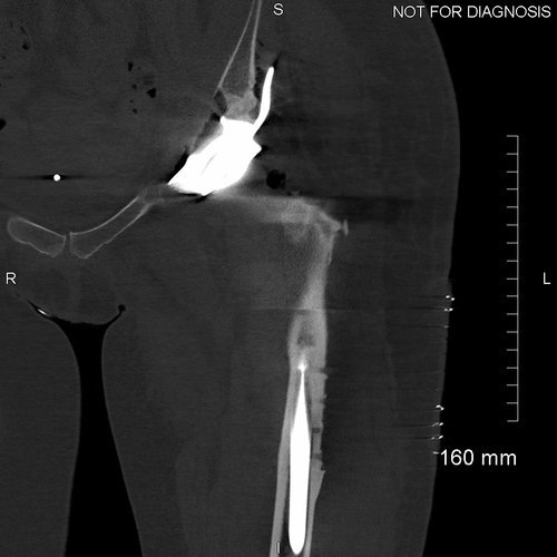
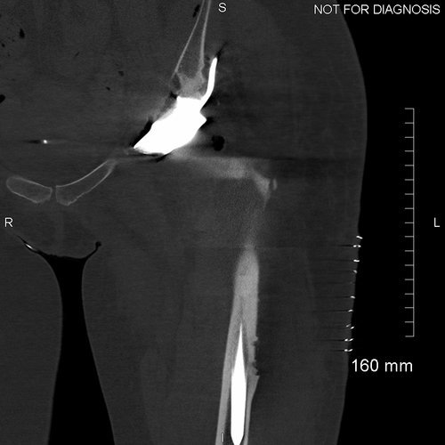
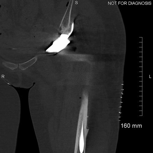
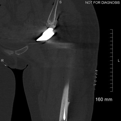
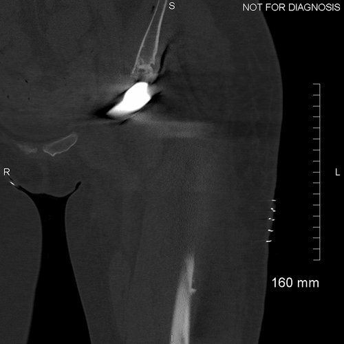
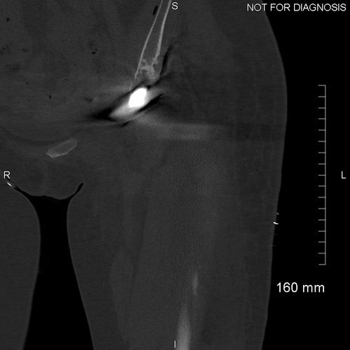
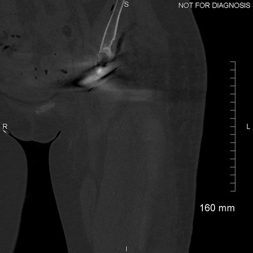
Coronal CT - This is used by the surgeon and the biomedical engineer to design the custom implant specifically to the patient’s acetabular bone stock. This CT demonstrates significant loss of bone stock relating to the left hip resurfacing.
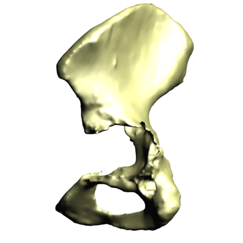
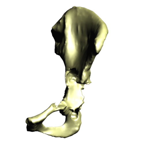
3D CT reconstruction showing the hemipelvic defect.
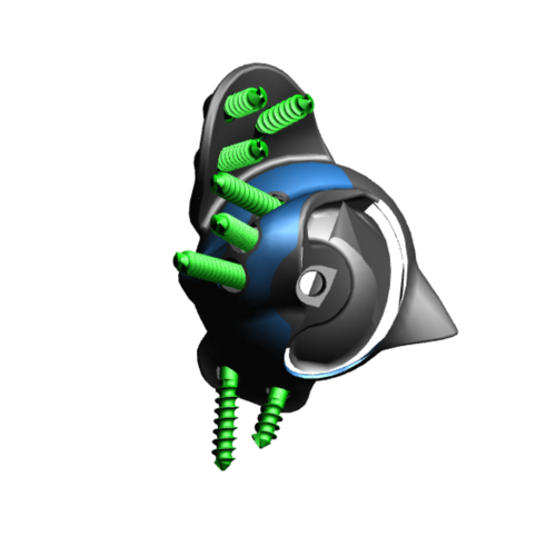
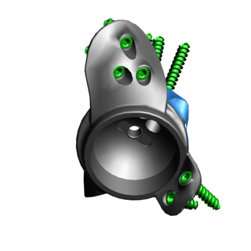
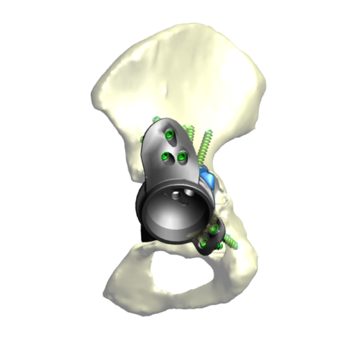
Implant design showing the custom acetabular implant designed around this patients bony anatomy. The blue areas highlight regions of trabecular titanium.
The Operation
We were able to use the old incision to remove the lateral plate from the femur. The femur was prepared for a modular, taper-fluted stem and Cup X explant was used to remove the cup.
We achieved minimal bone removal and reamed the acetabulum to the planned 60mm. The custom-made promade (3D-printed titanium acetabular component) fitted well with all screws, achieving good fixation and the planned length.
We fitted a dual mobility liner with ceramic head, checking that the leg length was good and the joint stable.
We washed the surgical site with Savlon and saline and then closed in layers with Ethibond, Vicryl and skin clips.
The Outcome
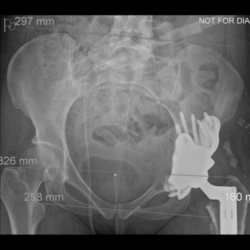
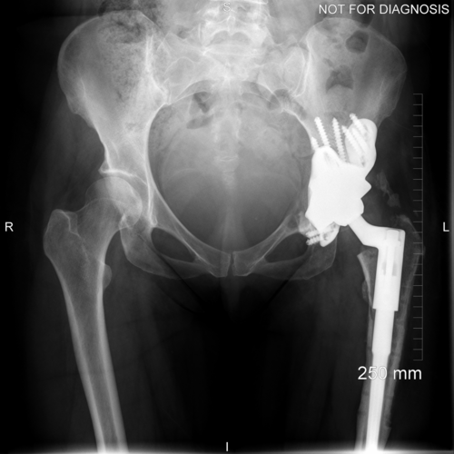
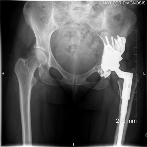
Anteroposterior plain radiograph showing the implant in situ with the surgical clips and a urinary catheter.


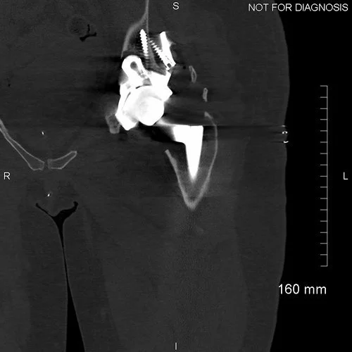


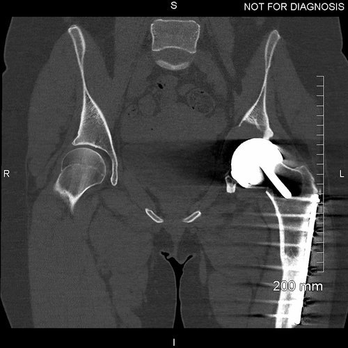
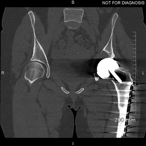
Coronal CT showing that the acetabular component is properly seated and the screws are within the bone.
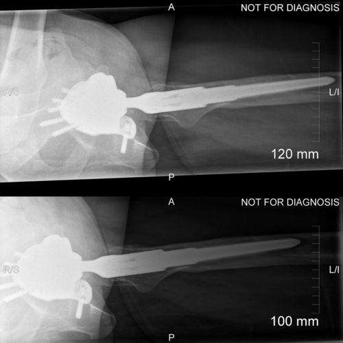
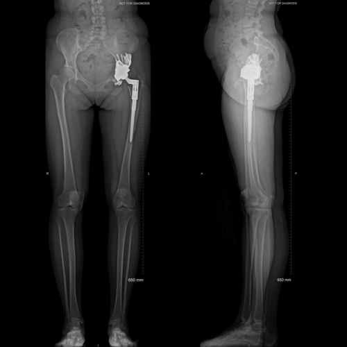
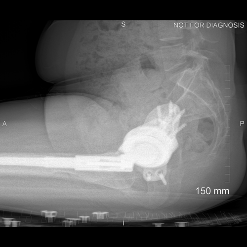
Anteroposterior and lateral plain radiographs taken 6-weeks after the operation. Zara was able to bear some weight on her leg with the use of two crutches. Her wound healed well and she was able to raise a straight leg.
EOS scanning taken 4-months after the operation. Zara was able to fully weight bear, she was trendelenburg negative and her EOS revealed no LLD. She had no pain in the joint and was using an exercise bike and swimming for rehabilitation.
Anteroposterior and lateral plain radiographs taken 6-months after the operation. Zara was able to walk without support; she had a good range of movement and was pain free. Her cobalt level was 11.3ppb and chromium level was 21.0ppb – a huge reduction.

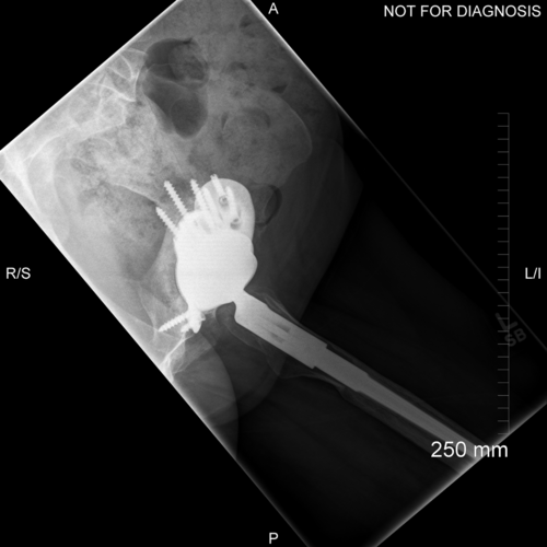
Anteroposterior plain radiograph taken at 1-year after the operation. There is no evidence of implant migration. Zara remained pain free and was very happy with the result of her new hip.
The Verdict
“The key to this case was the careful removal of the socket. There was a high risk of fracture during removal, which would have resulted in a very difficult fitting of the 3D-printed custom implant. The bone quality for fixation of a revision implant is uncertain when blood metal ion levels are 100 times greater than those of patients with well functioning MoM hips.
3D-printed custom implants increase the chance of fixation, particularly when there is minimal bone available and when the bone may have been poisoned by high levels of metal ions from MoM hips.”
-
Hart, A. J., Sabah, S., Henckel, J., Lewis, A., Cobb, J., Sampson, B., Mitchell, A. & Skinner, J. A. 2009. The painful metal-on-metal hip resurfacing. Journal of Bone and Joint Surgery-British Volume, 91B, 738-744.
-
-